Making sense of molecular structures at the atomic level
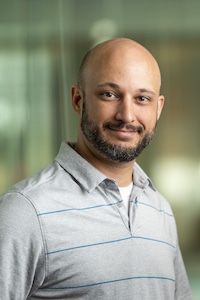
Peter Ducos made a military career of viewing the world from high above the earth, and a research career of examining the tiny particles of life up close.
As a U.S. Army soldier and government contractor instructor pilot of remotely operated aircraft, Ducos sat at a control panel not a lot different from the one he uses to control the technology involved in single-particle cryo-electron microscopy (cryo-EM).
“You have the joystick, and you have to make a map to navigate where you’re going, and then you collect information or data,” he says.
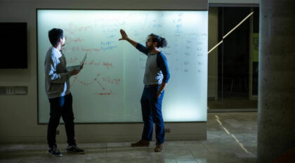
In his role as a research assistant, the biophysics doctoral student works in the Grant Lab, which collaborates with researchers across campus trying to answer complex and varied biological questions.

“The most rewarding aspect is when you get the initial view of what this stuff looks like, knowing no one’s ever seen this before,” says Ducos. “The first time you share the results with the people you’re working with, it’s almost like being a sonogram tech saying, ‘Here’s a picture of your baby,’ and you think they’re going to cry.”
Cryo-EM is one of the most promising technologies available to molecular biologists. The technique requires flash-freezing samples in liquid ethane that fixes proteins in glassy sheets of vitreous ice. From there, they can be examined and imaged using electron microscopes.
This revolutionary technology provides a breathtaking window into structural biology, allowing biological molecules, such as proteins, to be examined in three-dimensions through the generation of near-atomic reconstructions.
The models made possible by cryo-EM allow researchers to view the exquisite details of the molecules and gain insight into why functions may occur and the ability to predict interactions based on structure.
“If there’s something interesting going on, it makes it even better,” Ducos says. “It spurs entire sets of questions about what else we can test. What other structures can we look at? We’re not looking at individual molecules. It’s a collection of them and a snapshot in time. Then, we figure out how we get beyond that.”
After a five-year stint in the Army which took Ducos to places like Kosovo and Korea, he spent several years in Iraq as a government contractor. But the love of science that he harbored since childhood drew him to earn a bachelor’s degree in biology from the University of Missouri-Kansas City in 2019.
As an undergraduate, he switched majors from engineering to biology.
“When I made that decision, I decided I would make myself a learning aid and have some fun at the same time,” Ducos says. “I programmed a micro-controller for lighting and created a rainforest in a box.”
He obtained tropical plants and poison-dart frogs, bred fruit flies to feed the frogs, and introduced isopods and spring tails to act as clean-up crews in his mini-biome.
Ducos became interested in how the frogs’ toxins could have such drastic effects if they entered the bloodstream. Turns out, the toxin stops ion channels from allowing nerve cells to repolarize.
“As I started looking at structures, I thought these ion channels were really cool looking – they were literally the machines I was working with in engineering,” he says.
In the Grant lab, Ducos regularly leans on his military experiences.
“In the military, you learn to deal with all kinds of people working in a mission-oriented way,” says Ducos, who hopes to use his experience and Ph.D. to gain a government or private sector research job.
And whatever became of the poison-dart frogs? They continue to live happily in an expanded tropical environment in Ducos’ home office.

Rising Sparks: Early Career Stars
Rising Sparks is a monthly profile series exploring the personal inspirations and professional goals of early-career scientists at the Morgridge Institute.
