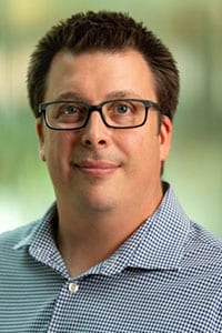
A new piece in the campus-wide puzzle of cryo-electron microscopy (or cryo-EM) is in place this month, as Tim Grant joins the investigator team of the Morgridge Institute for Research.
Grant, who joins Morgridge and UW–Madison from the Janelia Research Campus of the Howard Hughes Medical Institute (HHMI), is an expert on new methodologies to enhance images being generated from this game-changing technology. As an investigator at the institute’s John W. and Jeanne M. Rowe Center for Research on Virology, Grant will have the opportunity to partner with UW–Madison biologists to help perfect new tools that see deeper into cellular machinery.

Cryo-EM is a revolutionary imaging technology that allows biologists to see the structure of molecules within cells. Scientists can explore the very cellular surfaces where drugs and proteins interact, or where viruses orchestrate their cellular attacks. Simply, Cryo-EM could transform the development and delivery of precision medicine.
Bringing a project to life of this magnitude required almost a decade of work and roughly $16 million investment in technology and people. The new facility is expected to open this summer in the UW–Madison Biochemistry Building.
Grant says he is excited about the possibilities of this alliance between Morgridge and UW–Madison, as both entities will contribute to a fertile research environment. Grant’s faculty home will be in the Department of Biochemistry.
“From the UW–Madison side, the investment that has been made in cryo-EM is very impressive. And it highlights that there’s a commitment to it,” Grant says. “The equipment is great, but also the fact that the research is growing now and more and more people are getting involved.”
“There are also things that I want to do that are heavily computation-dependent,” he adds. “So from that side, the Morgridge Institute is a fantastic place to be, from the computational resources to the people who really know how to do large-scale computing on this level.”
Morgridge has a strong focus on research computing, and shares with UW–Madison the Center for High-Throughput Computing (CHTC), which allows scientists to vastly increase the size and scale of their research projects by harnessing distributed computing power.
Grant’s current research focus is addressing two challenges of current cryo-EM imaging. The first is being able to visualize smaller and smaller targets. There are entire classes of proteins now that cryo-EM can’t capture, he says.
” For me, the one dramatic impact will be extending things into a cellular context .”
Tim Grant
When scientists start pushing the limits on tiny structures, the only way to see them is with lower-resolution, less focused techniques, and they’re starting to hit a wall on what they can see and still glean meaningful data. Grant is developing a computational technique that marries both low- and high- resolution images within a single movie, enabling scientists to both see the structure and capture structural information.
Grant’s second focus second area is in capturing motion. Many biological structures are not static; they act more like machines with many moving parts.
“The great thing about those is, if you can capture those moving parts, not only do you open things up to solving new structures that couldn’t be solved before, but now you have this extra information about the motion,” he says. “So it not only solves a problem, it adds a whole layer of extra information.”
Grants says cryo-EM can contribute to an exciting future in cellular biology. It may help produce a complete molecular map of a cell, with all the topographic peaks and valleys where interactions take place.
“For me, the one dramatic impact will be extending things into a cellular context,” he says. “The idea will be to have potentially complete maps of exactly what is going on in the cell, where everything is in the cell, and how it’s working in the cell.
“It’s kind of like taking a step back and seeing the bigger picture,” he adds. “You know, right now we just look at isolated pieces. And that’s still very useful. But how those pieces interact is really more of a guessing game right now. Once you can move these things into a more cellular context, and see it directly, that will be very informative for how interactions occur between different things, and exactly how they work in real life.”
