Chimeric antigen receptor (CAR) T cell therapy is transforming cancer care for patients with cancers of the blood, but has proven especially challenging to develop against solid tumors.

Researchers at the Morgridge Institute and UW–Madison published new research in the journal Nature Biomedical Engineering using advanced metabolic imaging to better understand how cells function within the tumor microenvironment.
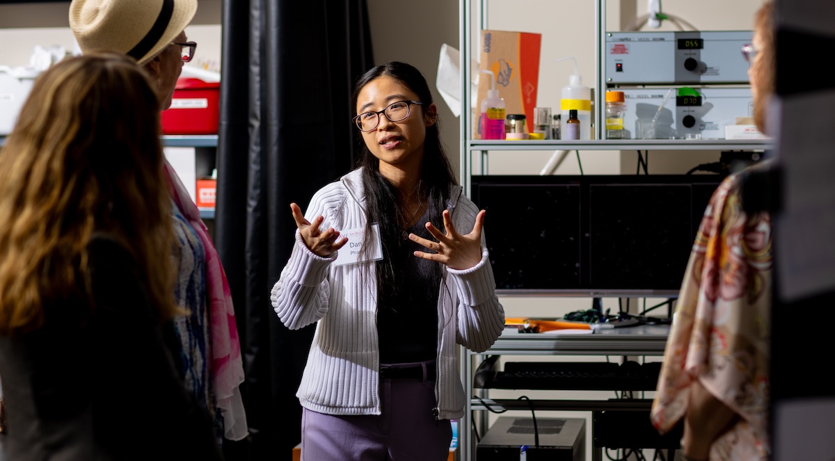
“Metabolism is one of the many factors that affects how T cells function within the tumor microenvironment, and hopefully one of the key factors that could inform what can be made better for these CAR T cells in a solid tumor,” says Dan Pham, a postdoctoral researcher in the lab of Morgridge Investigator Melissa Skala and first author of the study.
The tumor microenvironment is harsh and suppressive to immune cells with limited oxygen and glucose, both important elements for T cells to perform their function. However, during manufacturing, CAR T cells are grown in an artificial environment with unlimited nutrients and oxygen.
“When we put them in the body, we’re asking them, in a way, to survive in the wild,” says Skala. “Kind of like how you think of a captive lion being released into the wild — they don’t do so well.”
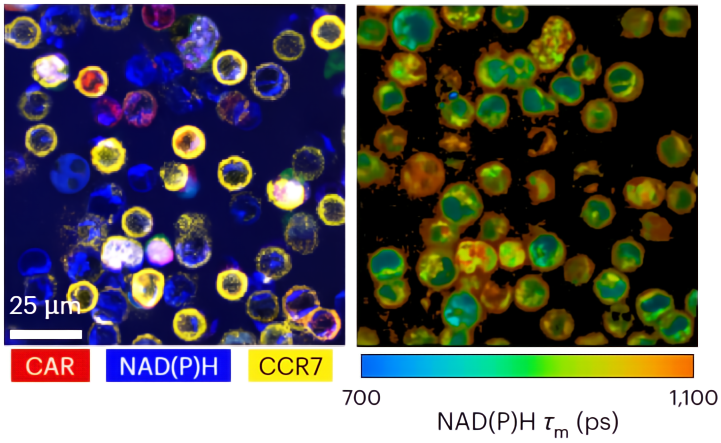
The Skala Lab uses a sensitive label-free imaging technique called optical metabolic imaging (OMI) to capture metabolic activity within the cells. A major benefit is being able to image a sample directly across many timepoints.

It also analyzes at the single-cell level, which can help differentiate populations that have stronger functional responses.
“This study hopefully supports the application of OMI to fill in the gaps in the analytical tools that people use for CAR T cell manufacturing,” says Pham. “Our measurements complement other metabolic assays from our collaborators so we can be certain in our validation.”
Collaborators include Morgridge metabolism investigator Jing Fan, UW pediatric oncologist and director of the Carbone Cancer Center Christian Capitini, and Wisconsin Institute for Discovery biomedical engineering investigator Kris Saha —who uses genomic editing tools like CRISPR CAS-9 to generate CAR T cells — and industry partner, Promega Corporation.
The researchers identified several windows of opportunity in the manufacturing process where optimization could result in a better therapeutic product, with a major focus on timing for gene editing.
“With OMI, we are able to sense when cells are entering the cell cycle, the optimal time for gene editing,” Pham says. “Over time, we can identify that peak when metabolism is supporting the cell cycle, and at that point they can do gene editing which would result in a higher percentage of CAR yield.”
Another area of optimization is in providing the right level of nutrients and metabolites at different manufacturing stages to enrich for the most functional T cells.
And at the end of the manufacturing process, OMI can help identify a product that would be more potent in vivo.
Pham says that this work is very much a proof of concept but ultimately demonstrates that OMI is sensitive enough to pinpoint these areas that could improve CAR T cell manufacturing. Since the technology is adaptable and scalable, there is potential to implement this tool in a large-scale manufacturing setting.
“There’s no point in building all this stuff unless you already know that you can impact the manufacturing process and predict which T cells will do better killing tumors in the body,” says Skala. “You have to establish all of that first, and once you do, then it’s worth building the technologies.”