When a patient is facing certain cancers or an immune condition, clinicians turn to their white blood cells for clues about the progression of both diseases and their treatments. From a standard blood draw, a clinical lab isolates peripheral blood mononuclear cells (PBMCs), which are predominantly immune cells, and then counts the abundance of different cell types, along with some basic measures of how they’re functioning.

“PBMCs can be isolated clinically really easily, and they’re already used in the clinical workflow,” says Melissa Skala, an investigator in biomedical engineering at the Morgridge Institute and senior author of a new study in Biophotonics Discovery. “So, the question is, what can we get from them that we aren’t already getting?”
Skala and her Morgridge Institute lab, including PhD student Jeremiah Riendeau, are working on bringing advanced imaging techniques to bear on the quest to retrieve as much information as possible from immune cells. In the process, they hope to help improve diagnosis and treatment of immune system-related conditions like blood cancers, lupus, sepsis, and cognitive decline. Additionally, PBMCs are the starting material for CAR T cell therapies, which engineer a patient’s own immune cells to fight certain cancers. Understanding immune dynamics in these engineered cells in more nuanced and comprehensive ways than are currently available to clinicians could improve the outcomes of patients undergoing treatments including CAR T cell therapy.
Previously, techniques to measure immune cell metabolism needed to isolate individual cell types or add chemical labels that highlight the presence or absence of metabolites. With the new study, Riendeau says the research team has, for the first time, accurately observed the metabolic state of single immune cells across a complex PBMC sample using a nondestructive analysis. When paired with current clinical techniques, this can provide a higher level of information beyond simple cell counts. Additionally, the classification of immune cells and their metabolic states in complex samples rather than monocultures provides more accurate insights into the cell’s behavior.
The ability to learn more about immune cells and their function without destroying them means the technique could be used in applications like assessing the fitness of immune cells before they’re administered as a treatment.

“You can continue using the samples after this analysis,” says Riendeau. With other techniques, “if you want to study metabolism you have to add different reagents to the sample, and that can be harmful to the cells.”
Skala adds that “a lot of starting material for cell therapies are PBMCs. If you could do something to assess the fitness of those cells before processing them for cell therapy, that would be nice.”
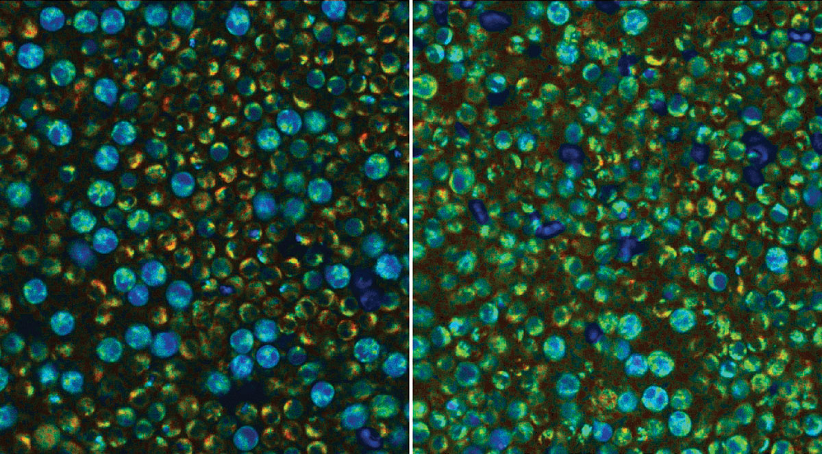
To achieve nondestructive and extremely high-resolution imaging, the team used a technique pioneered by the Skala Lab called optical metabolic imaging, or OMI, on samples of undifferentiated PBMCs isolated from donated blood of three human volunteers. With OMI, a sensitive microscope shoots two long-wavelength photons, which don’t damage material as much as shorter-wavelength ones but have the same effect, at the sample. This excites the inherent fluorescence of the products of cell metabolism and enables researchers to identify whether PBMCs are metabolically active or quiescent.
In this study, they did so with 93% accuracy only two hours after stimulating the cells. Further, they could specifically identify quiescent and activated monocytes with 96% and 88% accuracy, respectively, and natural killer (NK) cells in both states with 74% accuracy. While methods that use labels to identify immune cells and their metabolism are up to 100% accurate, the new technique has the distinct advantage of not needing to destructively manipulate the PBMCs. This makes OMI suitable for monitoring single-cell behavior over time while preserving samples for downstream applications.
Going forward, the team hopes to work with collaborators in hospitals to develop clinical applications for this analysis and bring better diagnosis and treatment outcomes to cancer and immune disorder patients.
“Not every lab has access to a two-photon microscope, and so part of the research that’s being done here is making this label-free metabolic information accessible to more labs,” says Riendeau. “Our lab and collaborators are in the process of commercializing our technology so we can make this measurement more available. The idea is, now that we understand how things look in a relevant sample, other labs can have this and do this, too.”
What are PBMCs?
PBMCs are any blood cell with a single nucleus and consist mostly of immune or white blood cells. This includes cells from both the innate and adaptive immune systems. Innate immune cells like NK cells and monocytes are nonspecific, meaning they respond to any foreign invaders they encounter, and can destroy infectious or cancerous cells before they cause problems. The adaptive immune system, including types of T and B cells, learn from past infections and target specific antigens on foreign cells.
“The innate immune cells,” says Riendeau, “are kind of like the first responders, and then later on the adaptive immune cells will come in and help out with a more target-specific approach.”
Why is immune metabolism so important?
The researchers found in the new study that innate immune cells are the most metabolically distinct, which likely is due to their ability to be highly reactive to threats and quickly ramp up their activity. Meanwhile, adaptive immune cells activate more slowly and need to be able to sustain energy over longer periods. But, elevated metabolic activity isn’t always good, and in conditions related to chronic inflammation, markers of activation can be an indicator that something is wrong.
“Generally, you don’t want to have constant, ongoing immune reactions. So, immune cells in the bloodstream are kept locked down in a quiescent state. But when there is some infection or other problem, like cancer, this would cause an immune reaction and activate metabolic pathways,” says Riendeau. “Having an understanding of the relative amounts of white blood cells is already used as a diagnostic biomarker. Adding this metabolic piece can tell you additional facts about the activation state of the immune cells.”
What can advanced imaging tools reveal about cell dynamics?
Riendeau says their new type of analysis using OMI can make sense of heterogenous metabolic states between cell types. For example, a subset of immune cells might be hyperactive while others remain quiescent. A bulk measurement averaging across the whole sample would hide those differences and obscure how the immune system is responding to an infection or whether cell therapy is successfully treating a cancer. But, with the ability to nondestructively measure metabolism of single immune cells in a complex sample, researchers will be able to push forward how we understand our body’s defense systems.