An expanding universe of collaboration
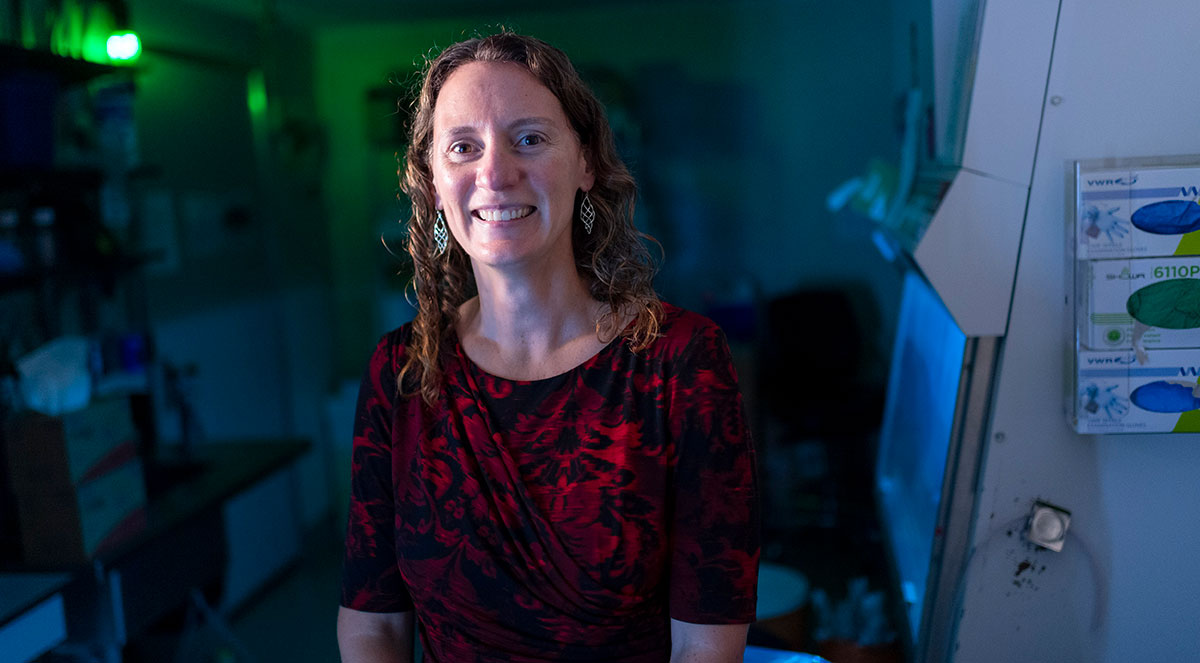
Melissa Skala quickly responds to an email from a collaborator before digging into a grant proposal with another. She’s stationed at a raised standing desk set up in her office — only she isn’t just standing, she’s walking on a portable treadmill tucked underneath.
“I’m very high energy, and I kind of have to use that energy to be able to focus,” she says enthusiastically.
Perhaps this energy is her secret to being a great scientific collaborator. It’s what drives her love for problem-solving. It powers her enthusiasm for championing others. And it’s infectious.
Over the past decade, the Skala Lab has become a powerhouse in the world of biomedical imaging. Skala has established collaborative relationships nationwide, working with researchers studying cancer, eye disease, maternal-fetal medicine, and more.
Underlying all these applications is a universe of cell biology that is always in motion. By pushing her lab’s signature imaging technologies to unveil its inner workings — without disrupting the system — the Skala Lab offers a transformative set of tools to biologists and clinicians alike. And with endless curiosity, she is fully charged to keep going.
Broadening the scope
Skala knew that she wanted to be a scientist in the first grade, when she developed a fascination with telescopes and their ability to reveal the secrets of the universe.
“I originally studied physics to be an astronomer,” she recalls from her early years of undergraduate studies at Washington State University. “I enjoyed working with my hands and building things.”
She eventually refocused her studies. Rather than using telescopic tools to observe the galaxies in the sky, she tinkered with lasers and light to build microscopy instruments that look at the galaxy within.
“There’s so much connection between astronomy and microscopy because of the technology, but we’re looking at cells and molecules in our bodies to see what we are made of, instead of looking at and beyond the universe,” she says.
Skala went on to complete her MS in Biomedical Engineering at UW–Madison and her PhD and postdoctoral training in Biomedical Engineering at Duke University. She then started her own lab at Vanderbilt University centered around her tool of choice: photons.
Pushing the boundaries of what is possible with photonics-based technologies, Skala’s research developed new ways to manipulate light and optics to better understand cancer and develop more effective therapies.
With a passion for big, fundamental research, Skala caught the attention of the Morgridge Institute for Research, a private biomedical research institute affiliated with UW–Madison. In 2016, she began her dual appointment as a Morgridge investigator and UW professor.
“We’re not metrics-driven at Morgridge. I have the privilege to be evaluated on if I’m doing good science,” Skala says. “I can take risks, think more deeply and hold on to a project until I think it’s ready to be published, which I think makes for better science and helps the scientific community more.”
Skala says that the partnership between the institute and the university helps advance science through building powerful research collaborations. It was this relationship that made the institute a particularly attractive place to establish her lab.
“It puts me in this great position to make connections between not only my lab, but other labs at Morgridge and UW. It just expands your network,” she adds. “Science is not done in isolation. You can do so much more if you partner with other people with expertise that is different than yours, but you have a similar goal. You ask more questions, get to learn more, and you just do better science.”
When approaching a project, Skala and her collaborators ask different questions. They want to know how to understand the biology of a system and she wants to know how to develop tools that can make things easier and better for them.
“I think it’s a good example of how to do science the right way — a way that benefits everyone and produces great results,” Skala says.
Of tools and trainees
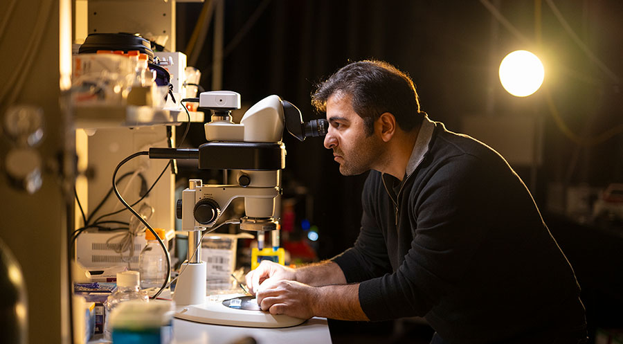
The Skala Lab works with a technique called fluorescence lifetime imaging microscopy (FLIM), specifically autofluorescence lifetime imaging. Instead of introducing fluorescent chemical tags to “label” biology of interest, the method uses naturally occurring fluorescence produced by cells when they are metabolically active.
The lab is best known for an innovative label-free technique called optical metabolic imaging (OMI). OMI uses two-photon microscopy to probe the fluorescence intensities and lifetimes of two metabolic co-enzymes, NAD(P)H and FAD. The intensity signals are used together to calculate the optical redox ratio — a way to quantify the metabolic changes and shifts in energy production and consumption within a cell. Calculating the fluorescence lifetimes of NAD(P)H and FAD gives additional insight into cell metabolism and functional states.
Since the method is non-invasive and non-toxic, it doesn’t damage living cells or tissues. This is a key benefit for biomedical research, where the ability to visualize living systems provides a critical advantage.
“It’s really exciting, because with her platform, we don’t have to perturb the cells,” says Dr. Christian Capitini, professor of pediatric hematology and oncology at UW–Madison School of Medicine and Public Health and a frequent collaborator with the Skala Lab. “We decrease the risk of contamination, and we also don’t waste any product for our assays.”
Early applications of OMI focused on methods to better understand cancer metabolism, particularly in breast cancer and pancreatic cancer. But over the years, Skala’s work became an attractive solution for biomedical researchers in various fields of expertise — neuroscience, eye disease, pre-term birth, and more.
“We’re making tools that can be used in very creative ways across disciplines and on many different problems, and I think that’s what makes them impactful,” says Skala. “And when you make them easier for others to use, the applications multiply and you don’t know where it’s going to go. That’s the fun part!”
As excited as she is when talking about technology and problem solving, she lights up even more when talking about being both a collaborator and mentor to those who come through her lab.
“The real impact is in the people I train and what they go on to do; I think that has 10 times more impact than any of the actual science that I’m doing in my lab,” she says. “I’m most excited seeing them succeed and learn and contribute in their own way.”
Skala cites the Morgridge Postdoctoral Fellowship program as a great boon to the collaborative nature of her work. Trainees are co-mentored by two principal investigators — one established at Morgridge and the other at UW–Madison. Less constrained by grant funding, these fellows have the flexibility to explore different disciplines that benefit from bridging research areas.
“I’ve learned the most about other areas of research through them,” she says. “It’s a great way to formalize a collaboration that you otherwise wouldn’t have initiated, because you need that person to introduce the idea.”
A case in point is Sabina Farhadova, the Carl E. Gulbrandsen Morgridge Postdoctoral Fellow. With Skala and Darcie Moore, associate professor of neuroscience at UW–Madison, Farhadova is applying optical metabolic imaging techniques to answer fundamental questions about stem cell biology and neurogenesis — the process by which new neurons are generated in the brain.
“The emphasis on collaboration, innovation and risk-taking creates an environment in which scientists can explore groundbreaking ideas without the constraints often found in traditional research settings,” Farhadova says.
One of the largest groups at the Morgridge Institute, the Skala Lab includes nearly 20 regular staff — including assistant scientists, technicians, postdoctoral researchers, and graduate students — plus a rotation of undergraduates and a few high schoolers throughout the school year.
Whether encouraging someone to follow their own career path into academia, industry, or somewhere in between, Skala offers the same advice: always keep your minds open to curiosity and collaboration.
Thinking like an engineer
Communication is vital in a good collaboration — and that can take time as engineers and biologists or clinicians learn each other’s language. Sean Palecek, a professor in chemical and biological engineering at UW–Madison, says he and Melissa found their shared language rather quickly.
“As engineers, we’re very process focused and not so much product focused,” he says. “And she has this great unique ability to match the technology with a problem and get excited by the applications as well as the technology.”
“I had a postdoc in my lab who came from Sean’s lab who started making me work on stem cells against my will. And now I’m like, ‘oh, I guess they’re okay,’” Skala says cheekily.
The Palecek Lab uses engineered microenvironments to study human pluripotent stem cells, which can differentiate into any cell type. Gaining a fundamental understanding of how these cells differentiate, develop, and mature will advance progress toward cell-based therapeutics and other clinical applications.
There are multiple factors that influence the fate of a cell, whether it is chemical, physical, or environmental. Thinking like an engineer, Palecek uses a systematic approach armed with a toolbox of multiple research strategies, including gene editing, tissue engineering, predictive modeling, multiomic data analysis, and now advanced imaging.
“I was really excited by Melissa’s skill set to be able to monitor metabolism, which is an important aspect of development — especially the heart, which is very metabolically active,” says Palecek. “And we really needed new technologies to help us track and characterize everything.”
The Palecek Lab developed several different protocols to differentiate stem cells into specialized heart cells, including cardiomyocytes, epicardial cells, smooth muscle cells and fibroblasts.
To completely characterize each of these cell types at every stage of differentiation would require nearly limitless resources. Standard methods of gathering those data require assays that are often time consuming, limited in scale, and use chemical reagents that often kill the cells.
“Right now, these stem cell differentiation processes are like following a recipe in a cookbook. You have your ingredients, you have your instructions, but you don’t do much characterization in the middle,” he says.
OMI offers a way to gather information collected over the cells’ lifetime, all without disrupting or damaging the cells. Palecek says this gives them a quick glance at the metabolic state of the cells, so if a batch is deviating from what they expect, they have a better fundamental understanding of why and how they might correct it to lead to different outcomes.
Skala says the approach is almost like working backwards — they know the final product should look like a batch of healthy and mature cells, so they need to figure out how to best monitor and control the process to achieve those results.
“The majority of people in science are hypothesis driven, but those of us who are engineers think about things in a fundamentally different way,” she says. “We’re asking ‘what are the tools you wish you had that you don’t know you need?’”
Palecek also serves as the deputy director for the NSF Engineering Research Center for Cell Manufacturing Technologies (CMaT), a national consortium that brings together collaborators from academia and industry working toward improving cell therapies. Their north star is to be able to design manufacturing methods that are simple, efficient, robust, reproducible and scalable.
“Almost all biomanufacturing these days uses this sort of monitoring and control, but cells aren’t there yet. And that’s where we’re trying to get the field,” he says.
Activating new science
When roaming the open common areas at Morgridge, it’s easy to find members of different labs congregating together for a casual lunch or briefly sharing their latest research finding. This interdisciplinary environment is by design.
Morgridge Investigator Jing Fan describes the workplace with an analogy fitting for a biochemist — it’s a giant catalyst for collaboration.
“You need to reach a point of activation energy for a reaction to occur. Being at an institute like this is basically catalyzing and bringing down that activation energy enough to spark a collaboration,” Fan says. “We’re exposed to different scientific questions and can learn, understand, and appreciate what others are doing and what technologies they are using.”
Fan came to the Morgridge Institute in 2017 with an office next door to Skala. She says right away, they were excited to chat each day and start building toward that activation energy.
A chemist and an engineer found their shared language in metabolism.
Like Skala, Fan’s early research initially focused on cancer metabolism, but her work expanded into the field of immunometabolism. The adaptive immune system uses T cells, which help establish immunological memory over the course of time. But the innate immune system requires cells like neutrophils or macrophages to act as first responders, rapidly warning the body of infection or disease.
“Innate immune cells are fascinating to me because they are very dynamic and heterogenous, even within their population they are metabolically different,” Fan says.
Techniques like mass spectrometry and isotope tracing provide detailed information about the biochemical mechanics of the molecules involved in cell function, but the cells are effectively destroyed after they are extracted and processed. Fan says learning more about OMI from the Skala Lab offered a way to better observe dynamic changes over time.
“The techniques they use and the approaches we use complement each other really well,” says Fan. “The imaging is really strong in that it uses live cells, it’s not destructive, and it has good resolution for bulk or single cells in different activated states.”
A short drive across campus, the Moore Lab is using OMI to study the inactive state of cells — neural stem cells, the precursor to “newborn” neurons that eventually mature and establish themselves in the circuitry of the brain.
With aging or disease, neural stem cells go into a deeper state of quiescence, or newly born neurons do not mature and die off, leading to cognitive decline. Moore says the regenerative process of stem cells in the brain is poorly understood due to limited tools that are sensitive enough to detect them in their dormant state.
“You can’t make newborn neurons if you can’t get the stem cells out of quiescence. If we can’t isolate these cells or identify them better, then we are not going to be able to understand how we can harness their power to start to make newborn neurons again,” she says.
In addition to reestablishing neuron pathways in the brain, neural stem cell regeneration could serve as a model for developing therapies for spinal cord injuries.
While the Moore Lab already had experience with several different imaging techniques, the collaboration with the Skala Lab pushed their technology, and the science, toward new applications.
“Darcie wanted to use our technology with a cell sorter so she could do single-cell RNA sequencing and relate it to what the cell will do later, if it has self-renewal capacity or not,” says Skala. “I never would have thought of that!”
Not unlike metabolism, great collaborations are dynamic as well, Moore says. The brainstorming phase is important, but you need that “spark” to turn ideas into action.
“When both people put the same energy into it, it’s very intoxicating to have really good chemistry with someone addressing different problems and for the science to move forward,” she says. “It feeds my scientific soul to have such a productive and exciting relationship where we can both say, ‘let’s try this crazy thing!’ and just do it.”
A bridge to better cell therapies
Traditional cancer therapies like radiation or chemotherapy are powerful options to combat cancer cells, but they also damage healthy cells in the body, resulting in undesired side effects.
There is great potential in chimeric antigen receptor therapy, or CAR T cell therapy, which uses a patient’s own immune cells — reprogramming the T cells to fight cancer cells. On the one hand, it is better tolerated by patients. But on the other hand, it takes time and resources to develop a personalized therapeutic.
“We work with Kris Saha, a CAR T cell expert, and Christian Capitini who is actually delivering cell therapies to cancer patients,” says Skala. “I’m never going to have the depth of expertise in those areas. I just know what a laser is and what it can do.”
Saha, a professor of biomedical engineering at UW–Madison and the Wisconsin Institute for Discovery, uses genomic editing tools like CRISPR-Cas9 to create CAR T cells that will specifically target cancer cells in a patient, leaving healthy cells alone.
Capitini, who recently became the director of the UW Carbone Cancer Center, treated patients with acute lymphoblastic leukemia at the UW Health American Family Children’s Hospital as part of the multi-site clinical trial that led to the first FDA-approved CAR T cell therapy in 2017. Since then, the FDA has approved only six other CAR T cell therapies for adults — all of them for blood cancers.
“Most cancer patients in the United States die from metastatic solid cancers, so that’s an area with a lot of potential for CAR T cell trials,” he says. “We’re looking at ways to weaponize the immune system to better recognize and fight targets on solid tumors.”
CAR T cell manufacturing technologies can take a small sample of cells to modify and expand to produce the millions of cells required for cell therapeutics. However, this rapid expansion makes the cells prone to exhaustion, says Capitini. The result is a mixture of some CAR T cells that are highly effective at targeting and killing cancer cells, but others in the batch will be less potent.
Saha and Capitini found that the CRISPR-based approach gives more precise control of modifying the cells, making them less prone to exhaustion and more efficient at recognizing cancers, including in solid tumors. “That gives them a higher chance of forming memory once it’s infused into people,” Capitini says.
Supported by CMaT, like the collaboration with Palecek, Skala is adapting OMI technology to help Saha and Capitini monitor and control CAR T cell production by measuring and comparing the metabolic state of the cells in real time.
“We want to know how we can identify more effective CAR T cells and promote their growth so they end up in the patient product and generate a better response,” she says.
Capitini also sees potential to take the technology from the pre-clinical side and make it a standard in the clinical practice. With personalized medicine like CAR T cell therapy, there is a benefit to real-time technologies that can help clinicians make a “go or no-go” decision on how to treat their patients.
Pre-clinical testing to fully characterize the CAR T cells includes methods like next-generation sequencing to gather data on genetic signatures that could help make predictions on treatment outcomes in the patient. But in a clinical setting, time is not often on their side.
“You have a patient waiting to receive therapy — you don’t want to wait two weeks until you have interpretable results — you want something that could be done the same day or next day and then make a decision,” he says. “We’re trying to figure out with this technology, can we get real-time information that is easily interpretable, and does it actually influence the outcome?”
For now, expertise in performing OMI techniques and data analysis is limited to the Skala Lab. But they are working to make their technologies more accessible and easier to use. Amani Gillette, an assistant scientist in the Skala Lab, recently founded SeLight, LLC, a startup company developing a portable imaging device to screen a patient’s cells before manufacturing.
“Our microscopes take up half a lab room, so we are engineering considerably smaller prototypes to get them out to clinics where doctors can use them,” Gillette says. “The more patients you can measure, the better understanding you have of what’s going on with a particular disease.”
With real-time imaging that is label-free, customizable, and low cost, Skala and Gillette hope their devices will prove to be a successful example of how academic research can translate into innovations that will advance healthcare.
Capitini shares that sentiment. While they are still in early stages of exploring the potential of live imaging for CAR T cell manufacturing, he hopes that through this collaboration, UW–Madison will eventually start using their own homegrown CAR T cell therapies.
“Melissa represents one of many investigators that are invested in improving these types of technologies,” he says. “It would be a new and exciting opportunity to really do this at scale and develop our own in-house workflows that incorporate her and others’ technologies and apply them to make cell therapy work better.”
The next frontier
Does scientific discovery necessitate new tools, or must tools be developed for scientific discovery to occur? For Skala, the answer is clear.
“I think discovery is empty without tools,” she says. “Discoveries can’t happen without new tools — it’s not possible.”
Tools are often seen as means to an end, says Skala, but it’s the tools that are opening doors and driving progress. Throughout history, major leaps of scientific discovery happened because of the tools that people used to explore the world around (and inside) them.
The first microscope was created in the late 16th century. Eighty years later, Antonie van Leeuwenhoek used a single-lens microscope to observe “tiny animals,” or single-celled microorganisms, in pond water. The first telescope was designed by a Dutch eyeglass maker as a magnification tool before Galileo Galilei pointed it to the sky to make astronomical observations. And NASA built the Voyager space probe to set out for the distant reaches of space, providing unexpected new information about the outer planets of our solar system along the way.
To Skala, science is a journey of discovery, and she sees imaging technology as the next frontier. What’s on the horizon? Higher resolution, greater specificity, faster speeds, computational models, and more.
“We’re just pushing the boundaries of what’s possible. There are a lot of opportunities for imaging, but it’s enormously complex,” she says. “If we can see smaller things, see faster things, see deeper things, we’re going to understand them better. We just don’t know how to do all that yet.”
With cutting-edge tools comes a higher level of expertise, which means fewer people who initially have the knowledge to use them. But for Skala, that’s what makes collaborative work so exciting.
“We ask, how are these tools useful? Then, how can we develop something that others can use? That’s our expertise,” she says. “It’s important to innovate. I think that’s what humans are good at, innovating and improving our lives. We enjoy it, and that’s why we do it. Bottom line.”
UW Partnership
Our strategic partnership with UW–Madison maximizes the impact of our research. Together we recruit top scientific talent, provide shared resources, and build powerful research collaborations like this one to keep Wisconsin science at the leading edge.
Learn more