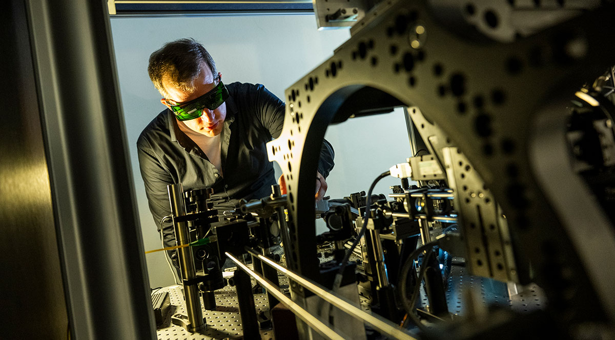
It’s easy for visitors to lose their bearings in the Laboratory for Optical and Computational Instrumentation (LOCI), a nondescript wing of the 54-year-old Animal Sciences Building at UW–Madison.
Thin hallways snake through more than a dozen small, interconnected labs, each one overstuffed with gleaming stainless-steel hardware, computer screens and black, wall-sized nylon partitions. Adding to the claustrophobic feel of the rooms is the near-complete absence of outside light.
The space certainly doesn’t give off the vibe of a high-tech wonderland. Yet for scientific imaging pioneers in Wisconsin, make no mistake: This is where the magic happens.

For Kevin Eliceiri, a professor of medical physics and biomedical engineering and inaugural biomedical imaging investigator at Morgridge, this space holds special meaning. He worked here as an undergraduate imaging assistant in the mid-1990s under the tutelage of two giants in modern cell biology, imaging pioneers Hans Ris and John White. Ris created the annex, now home to LOCI, to house a purpose-built three story million-volt High Voltage electron microscope to study cellular architecture. White built a prototype of a laser-scanning confocal microscope that became instrumental to modern biology.
Eliceiri continues this legacy of developing biological imaging instrumentation through his own work, yearly lectures named in honor of White and Ris, and a small microscopy museum documenting some of the history of microscopy at UW–Madison.
Six blocks from Discovery Building where Morgridge sits, LOCI serves as a “second campus” for instrument innovators at Morgridge who can stretch out and do their thing. As part of the Office of the Vice Chancellor for Research Center for Quantitative Cell Imaging (CQCI), directed by Eliceiri, it is the umbrella entity for the Eliceiri group and its sister biological optics lab run by Cell and Regenerative Biology Assistant Professor Abhishek Kumar. CQCI is a collaborative hub for biophotonics, the study of biology with light, at UW–Madison.
“One of the things that makes bioimaging so strong here is the breadth and depth across the board, because we’re a campus that has bought into the idea of strong arts and humanities, space sciences, physical sciences and biosciences, all working together,” Eliceiri says. “It allows imaging to thrive, because imaging fundamentally dips into all disciplines.”
That campus history helped fuel the biomedical imaging research theme at Morgridge, and the same interdisciplinary spirit served as the guiding force in hiring new investigators. Eliceiri describes ideal candidates as “the omnivores of the research world.”
Recruits have included biomedical engineer Melissa Skala, a leader in creating optical imaging technologies for use in cancer research and immunology; Randy Bartels, a physicist by training who is taking microscopy to new levels of depth and resolution; and data scientist Juan Caicedo, who is developing a digital atlas of cell behavior based upon billions of biological images.
“One thing I like about Morgridge is that we understand the power of imaging technology development for biology, which not everybody has traditionally invested in,” he says. “Some biologically focused centers would have been more conservative and said, ‘just hire a great cell biologist who uses imaging.’ But at Morgridge we also hired people that will take the imaging itself to new heights, and we’ve surrounded them with world-class biological faculty. We’ve been fairly unique in our goals to have co-localized experimental biologicals and imaging technologists.”
“It’s an interesting question: ‘Are the biologists driving us or are we driving them?’” Kevin Eliceiri
Eliceiri says this is a golden age for imaging. It began in the last 30-plus years with the digital revolution making staggering amounts of visual data available to science and now has the capabilities of deep learning and AI to extract greater biology insights from the images in ways that would be impossible for the human eye.
Morgridge serves as an amplifier of research potential and a hub for a lot of the expertise that exists in dozens of campus fields. Much like the Morgridge Metabolism Initiative, it is building community among like-minded scientists and seeding new directions.
“Imaging might be the most unifying element of all of Morgridge,” he says, noting that most of the 20-plus investigators at Morgridge have utilized or collaborated on improved imaging. “We have focused on hiring brilliant synthesizers between fields. It’s an interesting question: ‘Are the biologists driving us or are we driving them?’ The relationship is cyclical: biological needs inspire new imaging breakthroughs, and those breakthroughs open the door to unprecedented biological insights.”
One of the hallmarks of that collaboration is the Morgridge Fab Lab, which Eliceiri oversees. Located in the basement of Morgridge, it serves as a research-driven makerspace for all sorts of new ideas in biomedical imaging. That includes innovations in 3D printing; better visual diagnostics for cancer; improved transient lighting for surgery; and new instrumentation for multiscale imaging.
Another hallmark is BioImaging North America (BINA), an organization housed at Morgridge and supported by the Chan Zuckerberg Initiative (CZI). BINA is dedicated to helping biomedical imaging scientists better understand one another and the technologies they develop and utilize. It began in 2018 and has grown to more than 1,200 members from across the globe, including 200 core imaging facilities in the U.S., Mexico, and Canada.
Eliceiri has also been instrumental in improving and expanding an open-source imaging analysis software known as ImageJ. It’s an essential tool used by thousands of imaging scientists every day, valued for its ability to be customized for different fields and seamlessly plug in new capabilities like deep learning.
As a reflection of Morgridge’s innovative spirit, it is home to four distinct projects supported by CZI, which has a goal of supporting the science and technology that will make it possible to cure, manage or prevent all diseases by the end of the 21st century.
The future of imaging science in Madison is equally bright. Eliceiri organized a retreat this summer of two dozen UW–Madison scientists interested in building novel instruments for multiscale imaging. These developments could lead to novel imaging platforms for cell-informed medical imaging.
“We are at the cusp of fully appreciating the amazing phenomenon where everything good or bad in your life is happening within cells,” he says. “If you want to understand cells, you need novel technology to get to the dynamic fingerprints of the cell. We are well set up to do that.”
UW Partnership
Our strategic partnership with UW–Madison maximizes the impact of our research. Together we recruit top scientific talent, provide shared resources, and build powerful research collaborations like this one to keep Wisconsin science at the leading edge.
Learn more