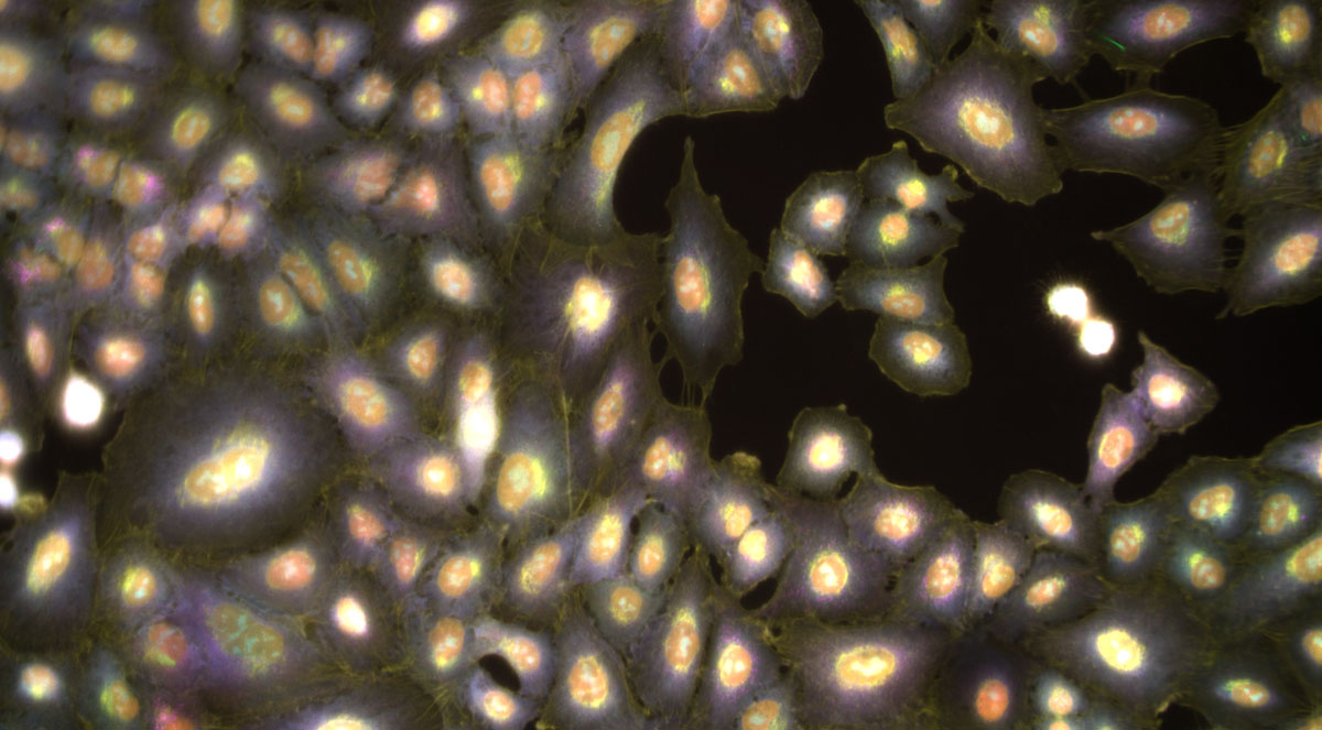
Anne Carpenter’s most significant research project was born out of frustration. In the late 1990s, Carpenter began a doctoral program in cell biology and started studying the structure of chromatin. The work required poring over microscopy images and painstakingly measuring the diameter of thousands of dots.
“This is terrible,” she remembers thinking. “There’s got to be a better way.”
Eventually she found image processing software called NIH Image (now known as ImageJ/Fiji, developed by Morgridge investigator Kevin Eliceiri) that allowed her to measure the slides in a quantitative and automated way. “I fell in absolute love,” she says. She had to tweak the program a bit to get it to do what she wanted, but once she got it working, “It was so much more satisfying,” she says.
Carpenter never dreamed she would go into computer sciences, but after her experience with NIH Image, she was hooked. She began writing her own software, an open-source program geared to the high-throughput experiments that she knew were coming. By 2005, she had drafted a program called CellProfiler that could easily locate the cellular features in a microscope image and measure various properties about them — shape, size, color, and pattern. In 2007, she established her lab at the Broad Institute of MIT and Harvard.
“The whole purpose is to — in a user-friendly and systematic way — help researchers extract the most information they can out of images.” CellProfiler can count a neuron’s synapses, measure the fat content of a worm, track cells as they move through time lapse images, count tumors in a mouse lung, and much, much more.
In academic science, success is often measured by the size of an individual’s lab. As the popularity of CellProfiler grew, so did Carpenter’s lab and, with it, her workload. In 2020, however, joint pain forced Carpenter to reevaluate the trajectory of her career. “I think scientists are often on autopilot. You just assume you should keep growing and getting as many grants and building your lab as large as possible,” she says. A doctor’s suggestion to reduce stress forced her to pause and think. “Where am I heading if I just keep going in this direction?”
Carpenter realized she wanted to stay closer to the science, not just oversee it as the head of a large lab. So she made an unconventional decision. She named Shantanu Singh, a computer scientist, as co-leader of her lab and transferred the future development of CellProfiler to staff scientist Beth Cimini, who was just launching her own group.
Cimini, who joined Carpenter’s lab as a postdoc in 2016, had been working on CellProfiler for years. She started using the software in graduate school and quickly became an evangelist. But there’s always room for improvement, and today Cimini spends much of her time trying to make image analysis even easier. “I spent a lot of years in graduate school doing things that felt like they were too hard,” she says. “I want to give people the knowledge to figure out how to make things work.”
For example, computers often struggle to define the boundaries of objects. “They rely basically on things either being dim at the edge or being round, and that’s not always how we see the boundaries of things.” But there are workarounds. Cimini recently helped Morgridge’s Melissa Skala, a biomedical engineer who works with images of cancer cells, find one. By incorporating a deep learning segmentation program called CellPose into CellProfiler, they figured out a better way to find the borders of these cells. “Now they don’t have to have all of these undergrads chained to desks for 10 hours a week circling cells,” she says.
With CellProfiler in good hands, Carpenter has been focused on a strategy she developed in 2013 called Cell Painting. In many microscopy experiments, biologists stain cells to highlight one feature that they expect to change in response to some perturbation. To Carpenter, that seemed like a missed opportunity. “You’re just cherry-picking this one thing that you happen to be researching,” she says. But “you have the potential to read out so many different things from every image.”
So Carpenter developed an assay to do just that. With Cell Painting, scientists douse the sample with six different dyes to highlight the cell’s most prominent organelles. This suite of simple stains can yield a surprising amount of information about the cells’ state and how different diseases, drugs, and genes affect them.
This is especially useful in drug discovery. If researchers expose cells to thousands of chemicals, for example, they can group them according to their effects. One group might be toxic to the liver. Another might be particularly effective for treating a disease. Pharmaceutical companies are already leveraging Carpenter’s tools to develop new therapies.
And Carpenter has her own drug discovery ambitions. In 2023, she co-founded SyzOnc, a startup that leverages Cell Painting and AI to develop therapies for hard-to-treat solid tumors.
Carpenter spent the first half of her career developing tools and methods. “I really hope to spend the next decades pushing forward different applications,” she says. “It feels to me like the time is right to really apply image analysis at large scale to accomplish great things for medicine.”
Editor’s note: Dr. Carpenter will be moving back to her Midwestern roots this summer, as her lab transitions to her undergraduate alma mater, Purdue University.